
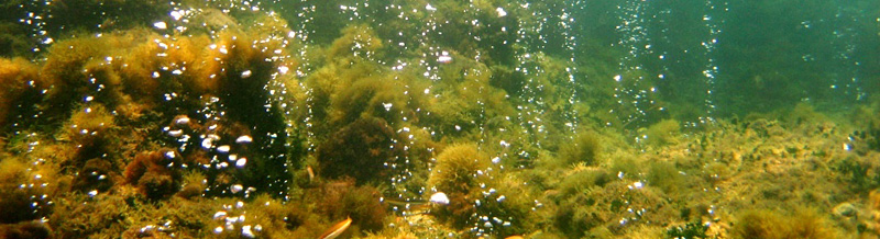
In 1898, Camillo Golgi described the intracellular structure that later took his name, the Golgi apparatus, which we now know to be involved in the transport and modification of proteins destined for secretion. The Golgi apparatus can be formed by single or multiple units; the latter can remain independent or connect to each other in a centralized structure called the Golgi 'ribbon'. The ribbon is generally considered an organization of the Golgi apparatus exclusively present in the cells of vertebrate animals. The reason why the Golgi ribbon has evolved, and its biological functions are not clear. However, in various pathologies, including neurodegenerative diseases, this organization of the Golgi apparatus is lost, indicating its importance for cellular physiology.
Working on cellular secretion, first at UConn Health in the United States and later at University College London in the United Kingdom, Dr. Ferraro has long been fascinated by the Golgi ribbon, hypothesizing that its functions can be deciphered through a comparative biology approach, from an evolutionary perspective. Back in Italy, at the Anton Dorhn Zoological Station, Dr. Ferraro focused his interests on the Golgi ribbon, conducting a study involving numerous colleagues from the Neapolitan institute and research centers in France, Spain, Germany, the United Kingdom, Norway, and the United States (*). The study, recently published in the journal Cell Reports, discovered that far from being exclusive to vertebrates, the Golgi ribbon is present in the cells of many taxonomic groups of animals. These observations indicate that this structure of the Golgi apparatus appeared early in the evolutionary history of animals, before their diversification into the groups existing today. The study also revealed that this structure, initially absent, forms during embryonic development in the sea urchin, ascidian, and amphioxus. This observation suggests the possibility that the Golgi ribbon has a function in embryonic development and that perhaps this is the ancestral role for which it evolved.
By revealing the unexpected and widespread presence of the Golgi ribbon among animals, the study, which relied on interdisciplinary collaboration of zoologists, cellular biologists, evolutionary biologists, and developmental biologists, brings the attention of the scientific community to this enigmatic structure and the importance of deciphering its functions. Future studies in this direction will allow for a better understanding of the role of the Golgi ribbon in the evolution of animal cells and in their physiology, and the consequences of its loss on the course of neurodegenerative diseases and other pathologies.
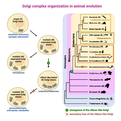
A morphological investigation of the Golgi apparatus in present-day species allowed the identification of the origin of the Golgi ribbon during the evolutionary history of animals in the common ancestor of cnidarians (jellyfish and corals) and bilaterians (all animals with bilateral symmetry).
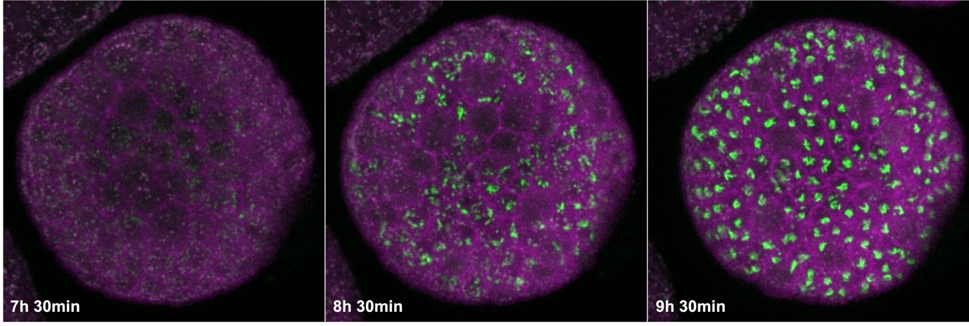
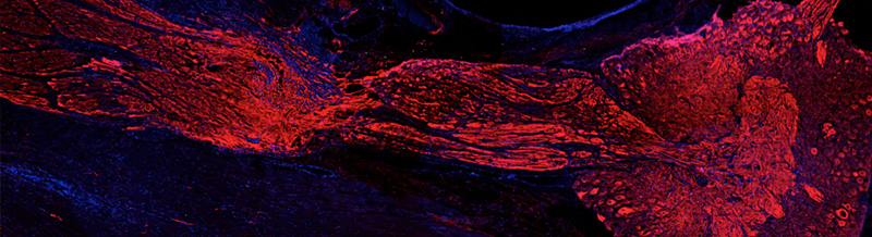
The images show a sea urchin embryo (Paracentrotus lividus) labeled with fluorescent reporters of the Golgi apparatus (green) and the plasma membrane (magenta). It can be observed how the Golgi apparatus, initially present as separate elements in embryonic cells, becomes a single structure, the Golgi ribbon. Times elapsed since fertilization are indicated.
Ream more:
(*) In addition to Francesco Ferraro, the researchers involved in the study were Giovanna Benvenuto, Serena Leone, Emanuele Astoricchio, Enrico D’Aniello, Salvatore D’Aniello, and Ina Arnone from the Anton Dohrn Zoological Station; Sophia Bormke, Jack Ullrich-Lüter, and Carsten Lüter from the Museum of Natural History in Berlin, Germany; Sanja Jasek and Gáspár Jékely from the Living Systems Institute at the University of Exeter in the United Kingdom; Maike Kittelmann from the Department of Biological and Medical Sciences at Oxford Brookes University in the United Kingdom; Kent McDonald from the University of California Berkeley in the United States; Volker Hartenstein from the Department of Molecular, Cell, and Developmental Biology at the University of California Los Angeles in the United States; Valentina Baena from the Department of Cell Biology at UConn Health in Farmington, United States; Héctor Escrivà and Stephanie Bertrand from the Institute of Integrative Biology of Marine Organisms at the Sorbonne University and CNRS in France; Bernd Schierwater from the Institute of Ecology and Evolution at the Hannover University of Veterinary Medicine Foundation in Germany; Pawel Burkhardt from the Michael Sars Centre at the University of Bergen in Norway; Iñaki Ruiz-Trillo from the Institute of Evolutionary Biology at Pompeu Fabra University in Barcelona, Spain. February 29, 2024 DOI:https://doi.org/10.1016/j.celrep.2024.113791