


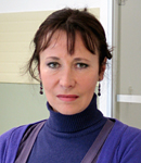
 Research Director
Research Infrastructures for Marine Biological Resources Department
Advanced Microscopy Center
Tel. +39 081 5833289 - +39 081 5833407 - +39 081 5833416
Fax: +39 081 7641355
e-mail luigia.santella(at)szn.it
Research Interests
Dr. Santella’s research has focused on the molecular mechanisms of signal transduction linked to the physiological changes in oocytes and eggs during meiotic maturation and fertilization, and in early embryos as well. Over the past 20 years, her research has placed special emphasis on the intracellular Ca2+ swings during these processes. Her work with starfish and sea urchin model systems has established that the Ca2+ wave in fertilized eggs can be recapitulated by combined effects of second messengers such as NAADP and InsP3 that play distinct roles in initiating and propagating the sperm-induced Ca2+ wave, while the other Ca2+-linked second messenger cADPr might instead have a modulatory role. She has also demonstrated that certain Ca2+ signals can originate and propagate in specific subcellular domains such as the nucleus and the subplasmalemmal regions. Her more recent work on starfish oocytes and eggs, which are optimally suited for microinjection and imaging analyses, has pioneered the new concept that the patterns of intracellular Ca2+ signaling and the Ca2+ ion channel activities are significantly modulated by the actin cytoskeleton. Dr. Santella has been active in promoting International collaboration and intellectual exchanges, as well as in stimulating education in foreign universities. She has also been involved in the organization of highly successful International symposia. She is regularly invited as a speaker at symposia and congresses in numerous countries in Europe, USA, and Asia. Since 1993, she has been a yearly visitor to the Asamushi Research Center for Marine Biology, Graduate School of Life Sciences, Tohoku University, Japan to perform seasonal work, and has lectured in the EMBO and NSF-sponsored educational programs in Brazil and Uruguay.
Selected Publications
Limatola N, Chun JT, Schneider SC, Schmitt JL, Lehn JM, Santella L. (2023) The Effect of Acidic and Alkaline Seawater on the F-Actin-Dependent Ca2+ Signals Following Insemination of Immature Starfish Oocytes and Mature Eggs. Cells 12 (5), 74
Santella L, Chun JT. (2022) Structural actin dynamics during oocyte maturation and fertilization. Biochem Biophys Res Commun 633, 13-16
Limatola N, Chun JT, Santella L. (2022) Species-Specific Gamete Interaction during Sea Urchin Fertilization: Roles of the Egg Jelly and Vitelline Layer. Cells 11 (19), 2984
Limatola N, Chun JT, Santella L. (2022) Regulation of the Actin Cytoskeleton-Linked Ca2+ Signaling by Intracellular pH in Fertilized Eggs of Sea Urchin. Cells 11 (9), 1496
Limatola N, Chun JT, Cherraben S, Schmitt JL, Lehn JM, Santella L. (2021) Effects of dithiothreitol on fertilization and early development in sea urchin. Cells 10 (12), 3573
Limatola N, Vasilev F, Santella L, Chun JT. (2020) Nicotine induces polyspermy in sea urchin eggs through a non-cholinergic pathway modulating actin dynamics. Cells, 9, 63
Santella L, Limatola N, Chun JT. (2020) Cellular and molecular aspects of oocyte maturation and fertilization: a perspective from the actin cytoskeleton. Zool Lett., 6, 1-21
Limatola N, Chun JT & Santella L. (2020) Effects of salinity and pH of seawater on the reproduction of the sea urchin Paracentrotus lividus. Biol Bull., 239, 13-23
Limatola N, Vasilev F, Chun JT & Santella L. (2019) Sodium-mediated fast electrical depolarization does not prevent polyspermic fertilization in Paracentrotus lividus eggs. Zygote, 27, 241-24
Vasilev F, Limatola N, Chun JT & Santella L. (2019) Contributions of suboolemmal acidic vesicles and microvilli to the intracellular Ca2+ increase in the sea urchin eggs at fertilization. Int J Biol Sci., 15, 757
Limatola N, Vasilev F, Chun JT & Santella L. (2019) Altered actin cytoskeleton in ageing eggs of starfish affects fertilization process. Exp Cell Res., 381, 179-190
Limatola N, Chun JT, Kyozuka K, Santella L. (2015) Novel Ca2+ increases in the maturing oocytes of starfish during the germinal vesicle breakdown. Cell Calcium, 58, 500-510
Chun JT, Limatola N, Vasilev F, Santella L. (2014) Early events of fertilization in sea urchin eggs are sensitive to actin-binding organic molecules. Biochem Biophys Res Commun., 450, 1166-74
Vasilev F, Chun JT, Gragnaniello G, Garante E, Santella L. (2012). Effects of ionomycin on egg activation and early development in starfish. PLoS One, 7, e39231
Chun JT, Puppo A, Vasilev F, Gragnaniello G, Garante E, Santella L. (2010) The biphasic increase of PIP2 in the fertilized eggs of starfish: new roles in actin polymerization and Ca2+ signaling. PLoS One, 5, e14100
Puppo A, Chun JT, Gragnaniello G, Garante E, Santella L. (2008). Alteration of the cortical actin cytoskeleton deregulates Ca2+ signaling, monospermic fertilization, and sperm entry. PLoS One 3, e3588
Kyozuka K, Chun JT, Puppo A, Gragnaniello G, Garante E, Santella L. (2008). Actin cytoskeleton modulates calcium signaling during maturation of starfish oocytes. Dev Biol., 320, 426-35
Lim D, Ercolano E, Kyozuka K, Nusco GA, Moccia F, Lange K, Santella L. (2003) The M-phase-promoting factor modulates the sensitivity of the Ca2+ stores to inositol 1, 4, 5-trisphosphate via the actin cytoskeleton. J Biol Chem., 278, 42505-42514
Lim D, Lange K, Santella L. (2002) Activation of oocytes by latrunculin A. The FASEB J., 16, 1050-1056
Lim D, Kyozuka K, Gragnaniello G, Carafoli E, Santella L. (2001) NAADP+ initiates the Ca2+ response during fertilization of starfish oocytes. The FASEB J., 15, 2257-226
Santella L, Kyozuka K. (1994) Reinitiation of meiosis in starfish oocytes requires an increase in nuclear Ca2+. Biochem Biophys Res Commun., 203, 674-680
Websites or online articles
The fertilization process: a new way to look at an old phenomenon
2015 Small World in Motion Competitionn (Video)
International Symposium - The dynamically active egg: The legacy of Ernest Everett Just
Byrnes W M. (2013). Walking in the Footsteps of Ernest Everett Just at the Stazione Zoologica in Naples: Celebration of a Friendship
Photos
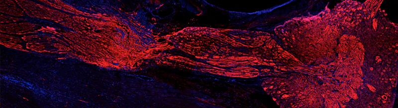
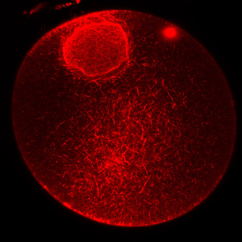
F-actin staining in a starfish oocyte microinjected with Alexa Fluor-568 conjugated phalloidin.
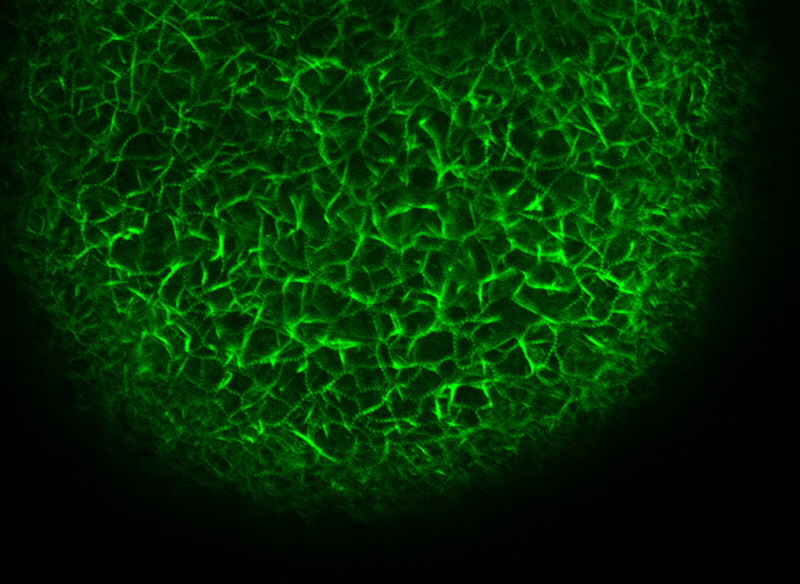
F-actin staining in the nucleus and cytoplasm of a starfish immature oocyte.
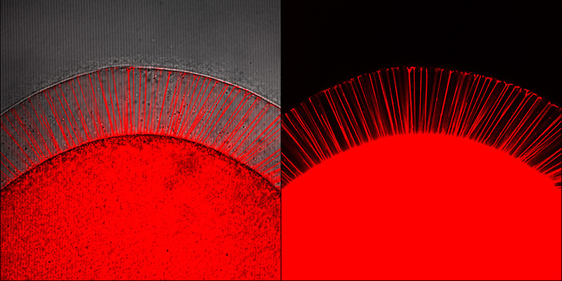
F-actin spikes into the perivitelline space of a starfish egg following uncaging of injected InsP3 visualized by labelling PIP2 with the RFP-PH domain of the PLC1.
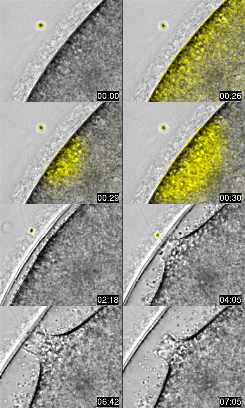
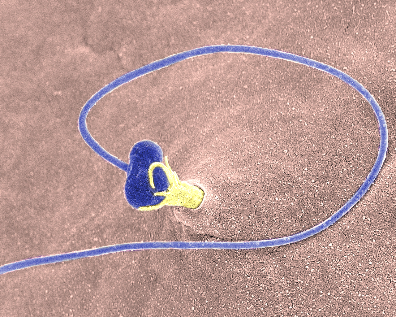
The interaction of the acrosomal process, visible at 04:05 min, of the sperm (yellow circle) with the jelly coat of a starfish egg triggers intracellular Ca2+ increases (yellow fluorescence). Detailed view of the sperm entry site during fertilization.
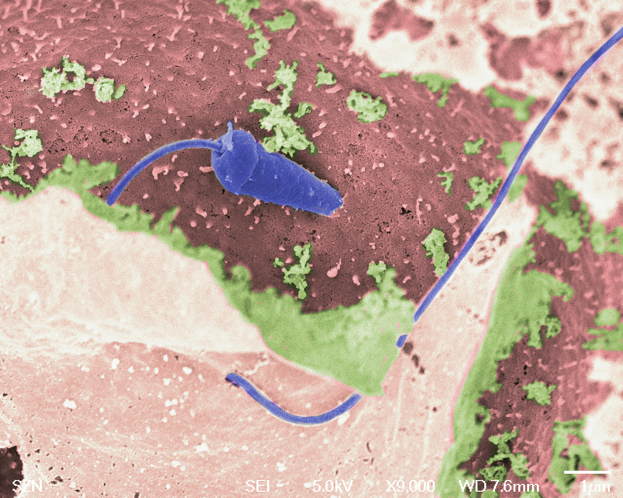
Sperm incorporation into a starfish egg by cytoplasmic protrusions crossing the fertilization envelope.
Starfish bipinnaria larvae three days after fertilization.
Sperm-egg fusion beneath the fertilization envelope following insemination of a sea urchin egg.
Copyright © 2015 by Stazione Zoologica Anton Dohrn Napoli - Italy
Villa Comunale, 80121 Napoli - Tel.+39 081 5833111 - C.F./P.IVA : IT 04894530635 - PEC: ufficio.protocollo(at)cert.szn.it